SCIENCE AND TECHNOLOGY
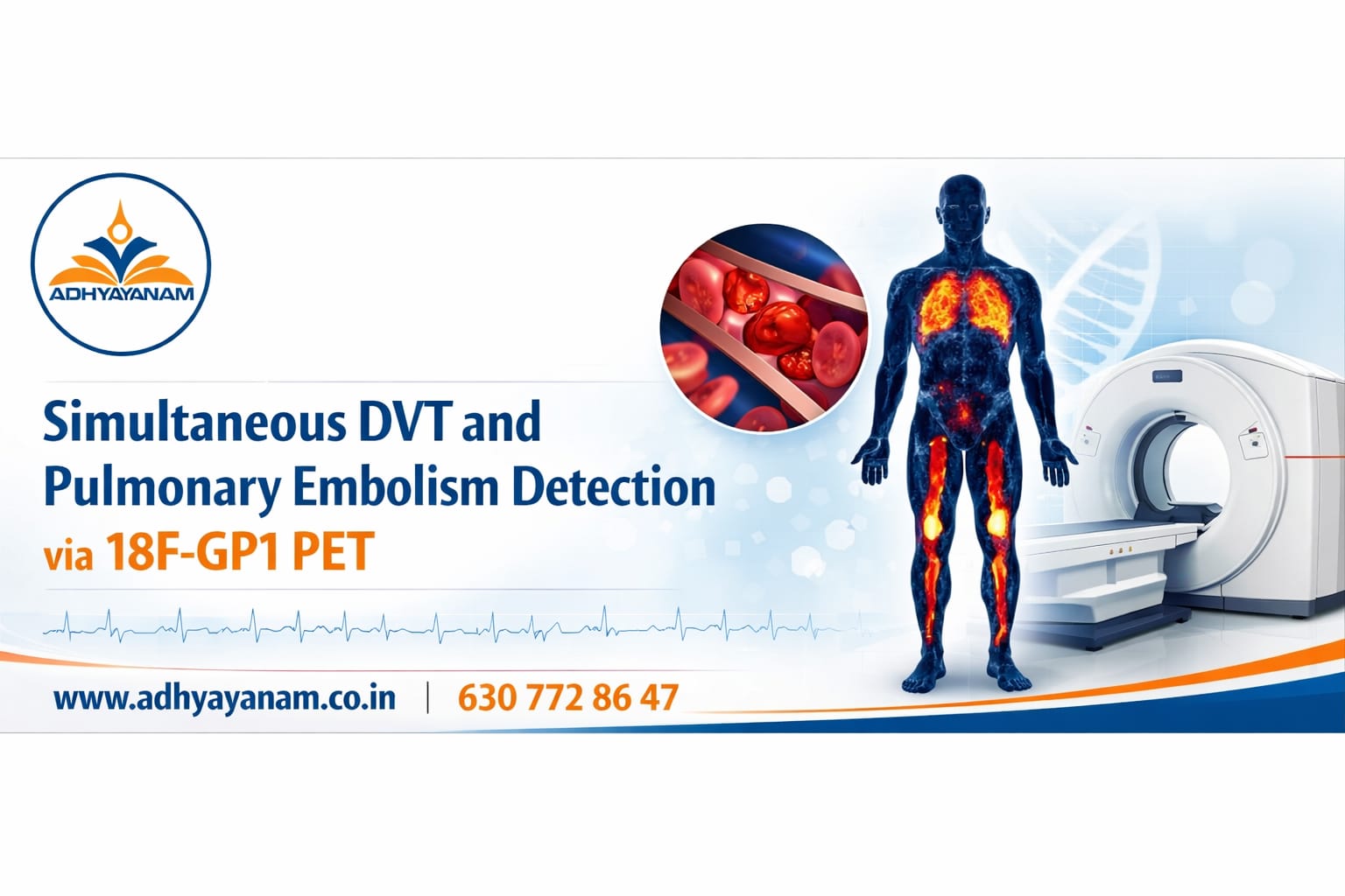
Simultaneous DVT and Pulmonary Embolism Detection via 18F-GP1 PET
The medical community is celebrating a major leap forward in vascular diagnostics as a groundbreaking PET radiotracer was recently named “Image of the Year” at the Society of Nuclear Medicine and Molecular Imaging (SNMMI) 2026 annual meeting. Developed by Dr. Sangwon Han and his team at the Asan Medical Centre in Korea, this innovative technology allows for the first time a single, comprehensive scan that can visualize blood clots throughout the entire body.
Addressing a Silent Killer
Deep vein thrombosis (DVT)—clots typically forming in the deep veins of the legs—is a prevalent condition with an incidence rate nearly half that of all cancers combined. The primary danger of DVT is its potential to develop into a pulmonary embolism, a life-threatening event where a clot breaks loose and blocks blood flow to the lungs. Because early detection is the primary factor in determining patient treatment and survival, the need for precise imaging has never been higher.
The Limits of Traditional Imaging
Historically, clinicians have relied on venous ultrasonography (VUS) or CT scans to diagnose these conditions. However, these methods have significant drawbacks. VUS is highly dependent on the skill of the operator, struggles to detect clots in the calf, and often relies on indirect structural changes rather than seeing the clot itself.
In contrast, the new tracer, known as fluorinated GP1 (18F-GP1), is "thrombus-targeted". It specifically binds to receptors on activated platelets, the actual cell fragments responsible for forming clots. This allows physicians to directly visualize active thrombus formation anywhere in the body.
High Accuracy and Comprehensive Detection
In a Phase 2 clinical study involving 46 symptomatic patients, the 18F-GP1 PET/CT demonstrated remarkable diagnostic power. The researchers performed chest-to-feet scans approximately two hours after administering the tracer. The results revealed a 95% sensitivity and 92% specificity for detecting proximal DVT (clots in the pelvic, thigh, or knee regions).
Furthermore, the technology showed strong agreement with traditional methods for distal (calf) clots and displayed excellent reproducibility among different readers. One of the most significant advantages is the ability to identify concomitant pulmonary emboli during the same session used to check the legs, streamlining the diagnostic process for critically ill patients.
A New Frontier in Nuclear Medicine
The award-winning image captured by Dr. Han’s team revealed extensive clots not only in expected areas like the legs and lungs but also in unusual sites such as cranial vessels, spinal vessels, and cardiac valves. This level of detail was achieved with no drug-related adverse events, suggesting the tracer is both safe and well-tolerated.
Looking ahead, the potential for 18F-GP1 extends far beyond DVT. This technology represents a vital step toward thrombus-specific imaging that could revolutionize the treatment of embolic strokes and various other cardiovascular diseases. By providing a clear, whole-body map of active clotting, this tool is set to become a cornerstone of future nuclear medicine.
SOURCE : PHYSICS WORLD

OTP